

Article Sidebar
Main Article Content
Abstract
Background: Non-invasive cardiac imaging is increasingly used nowadays to predict and diagnose structural and functional diseases of coronary arteries with improved efficacy, safety and cost.
Objective: To evaluate the ability of two-dimensional (2D) global longitudinal strain (GLS) by speckle echocardiography to predict the presence of significantcoronary artery disease (CAD), disease severity and its’ reproducibility in the stratification of coronary disease risk in suspected patients.
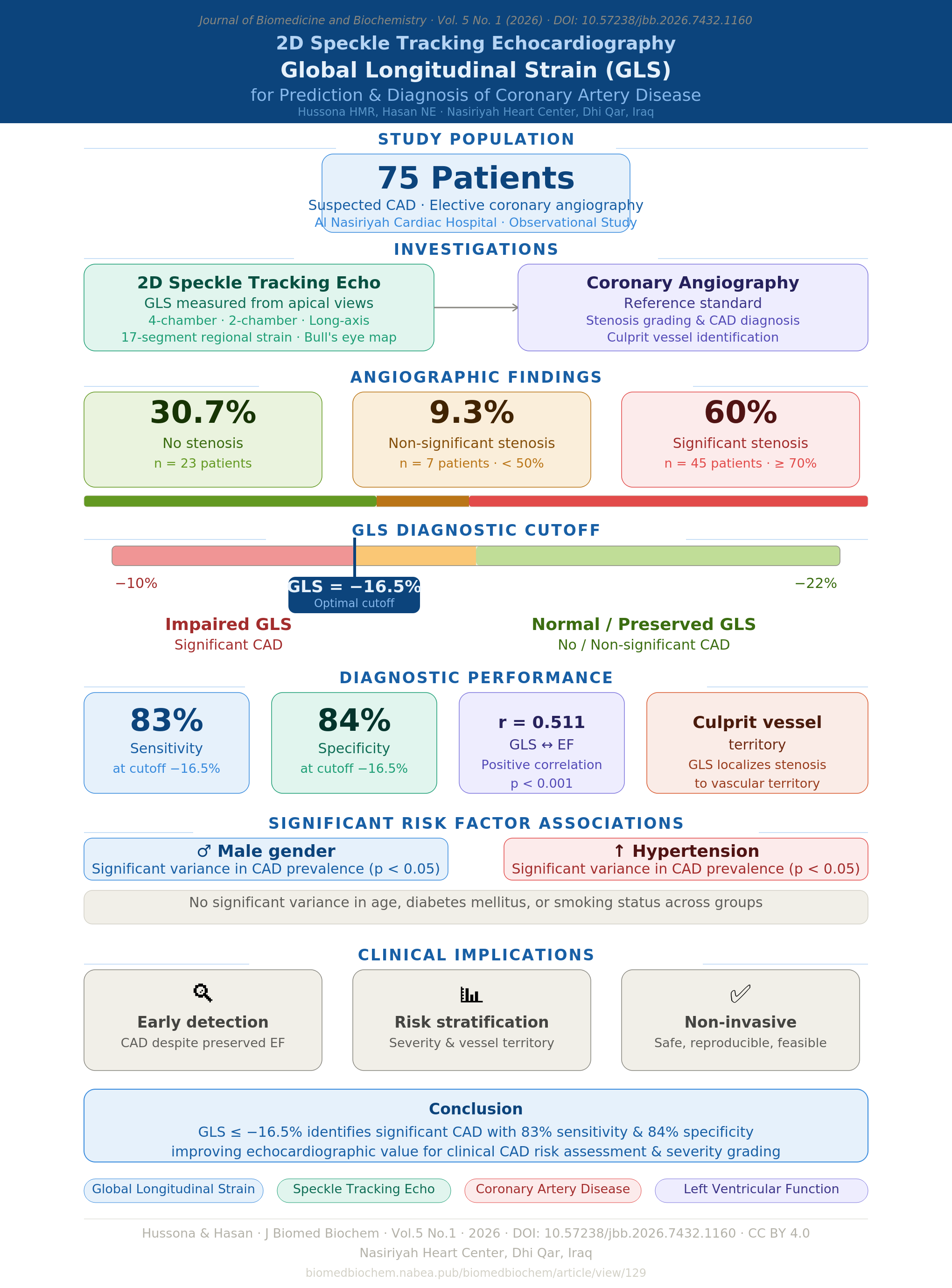
Patients and methods: An observational study that was conducted at Al Nasiriya Cardiac Hospital and included 75 patients with suspected CAD who were admitted for elective diagnostic coronary angiography. They divided into 3 groups according to presence of coronary artery stenosis andthe degree of significance. 2D speckle tracking echocardiography was performed for all and its’ ability in the prediction and diagnosis of CAD was studied.
Results: Study showed that 60% of patients had significant stenosis, while 30.67% had no stenosis and 9.33% had a non- significant stenosis by coronary angiography. There was a significant variance regarding gender preference and hypertension with no significant variance regarding age,diabetes and smoking status. GLS had significant associations with the degree of coronary stenosis and culprit vessels, having a cut of value of -16.5% for discrimination of significant, non-significant and no stenosis with 83% sensitivity and 84% specificity. GLS had a significant positivecorrelation with the ejection fraction, r = 0.511 at a p-value <0.001.
Conclusions: 2D speckle tracking echocardiogram GLS has the potential to improve the value of echocardiography in identifying high-riskpatients, detecting CAD and CAD severity (degree of stenosis) which provides more information for the clinical physician.
Keywords
Article Details
Copyright (c) 2026 Hyder Mejeed Rustum Hussona, Nazar Essa Hasan (Author)

This work is licensed under a Creative Commons Attribution 4.0 International License.
How to Cite


References
- Patel MR, Peterson ED, Dai D, Brennan JM, Redberg RF, Anderson HV, et al. Low diagnostic yield of elective coronary angiography. N Engl J Med. 2010;362(10):886-895. https://doi.org/10.1056/NEJMoa0907272
- Medvedofsky D, Kebed K, Laffin L, et al. Reproducibility and experience dependence of echocardiographic indices of left ventricular function: side-by-side comparison of global longitudinal strain and ejection fraction. Echocardiography. 2017;34(3):365-370. https://doi.org/10.1111/echo.13457
- Radwan H, Hussein E. Value of global longitudinal strain by two-dimensional speckle tracking echocardiography in predicting coronary artery disease severity. Egypt Heart J. 2017;69(2):95-101. https://doi.org/10.1016/j.ehj.2016.11.002
- Hoit BD. Strain and strain rate echocardiography and coronary artery disease. Circ Cardiovasc Imaging. 2011;4(2):179-190. https://doi.org/10.1161/CIRCIMAGING.110.960872
- Choi SW, Park JH, Sun BJ, et al. Impaired two-dimensional global longitudinal strain of left ventricle predicts adverse long-term clinical outcomes in patients with acute myocardial infarction. Int J Cardiol. 2015;196:165-167. https://doi.org/10.1016/j.ijcard.2015.05.133
- Kearney LG, Lu K, Ord M, et al. Global longitudinal strain is a strong independent predictor of all-cause mortality in patients with aortic stenosis. Eur Heart J Cardiovasc Imaging. 2012;13(10):827-833. https://doi.org/10.1093/ehjci/jes115
- Kusunose K, Goodman A, Parikh R, et al. Incremental prognostic value of left ventricular global longitudinal strain in patients with aortic stenosis and preserved ejection fraction. Circ Cardiovasc Imaging. 2014;7(6):938-945. https://doi.org/10.1161/CIRCIMAGING.114.002041
- Saito M, Negishi K, Eskandari M, et al. Association of left ventricular strain with 30-day mortality and readmission in patients with heart failure. J Am Soc Echocardiogr. 2015;28(6):652-666. https://doi.org/10.1016/j.echo.2015.02.016
- Potter E, Marwick TH. Assessment of left ventricular function by echocardiography: the case for routinely adding global longitudinal strain to ejection fraction. JACC Cardiovasc Imaging. 2018;11(2 Pt 1):260-274. https://doi.org/10.1016/j.jcmg.2017.11.017
- Parato VM, Mehta A, Delfino D, et al. Resting echocardiography for the early detection of acute coronary syndromes in chest pain unit patients. Echocardiography. 2010;27(5):597-602. https://doi.org/10.1111/j.1540-8175.2010.01166.x
- Reant P, Labrousse L, Lafitte S, et al. Experimental validation of circumferential, longitudinal, and radial 2-dimensional strain during dobutamine stress echocardiography in ischemic conditions. J Am Coll Cardiol. 2008;51(2):149-157. https://doi.org/10.1016/j.jacc.2007.08.039
- Yingchoncharoen T, Agarwal S, Popovic ZB, Marwick TH. Normal ranges of left ventricular strain: a meta-analysis. J Am Soc Echocardiogr. 2013;26(2):185-191. https://doi.org/10.1016/j.echo.2012.10.008
- Yang ZR, Zhou QC, Lee L, et al. Quantitative assessment of left ventricular systolic function in patients with coronary heart disease by velocity vector imaging. Echocardiography. 2012;29(3):340-345. https://doi.org/10.1111/j.1540-8175.2011.01578.x
- Belghitia H, Brette S, Lafitte S, et al. Automated function imaging: a new operator-independent strain method for assessing left ventricular function. Arch Cardiovasc Dis. 2008;101(3):163-169. https://doi.org/10.1016/j.acvd.2008.02.003
- Nucifora G, Schuijf JD, Delgado V, Bertini M, Scholte AJ, Ng AC, et al. Incremental value of subclinical left ventricular systolic dysfunction for the identification of patients with obstructive coronary artery disease. Am Heart J. 2010;159(1):148-157. https://doi.org/10.1016/j.ahj.2009.10.030
- Shimoni S, Gendelmann G, Ayzenberg O, et al. Differential effects of coronary artery stenosis on myocardial function: the value of myocardial strain analysis for the detection of coronary artery disease. J Am Soc Echocardiogr. 2011;24(7):748-757. https://doi.org/10.1016/j.echo.2011.03.007
- Montgomery DE, Puthumana JJ, Fox JM, Ogunyankin KO. Global longitudinal strain aids the detection of non-obstructive coronary artery disease in the resting echocardiogram. Eur Heart J Cardiovasc Imaging. 2012;13(7):579-587. https://doi.org/10.1093/ejechocard/jer282
- Smedsrud MK, Sarvari S, Haugaa KH, Gjesdal O, Orn S, Aaberge L, et al. Duration of myocardial early systolic lengthening predicts the presence of significant coronary artery disease. J Am Coll Cardiol. 2012;60(12):1086-1093. https://doi.org/10.1016/j.jacc.2012.06.022
- Biering-Sørensen T, Hoffman S, Mogelvang R, et al. Myocardial strain analysis by 2-dimensional speckle tracking echocardiography improves diagnostics of coronary artery stenosis in stable angina pectoris. Circ Cardiovasc Imaging. 2014;7(1):58-65. https://doi.org/10.1161/CIRCIMAGING.113.000989
- Evensen K, Sarvari S, Ronning M, et al. Carotid artery intima-media thickness is closely related to impaired left ventricular function in patients with coronary artery disease: a single-centre, blinded, non-randomized study. Cardiovasc Ultrasound. 2014;12:39. https://doi.org/10.1186/1476-7120-12-39
- Billehaug N, Edvardsen T, et al. Diagnostic accuracy of left ventricular longitudinal function by speckle tracking echocardiography to predict significant coronary artery stenosis. BMC Med Imaging. 2015;15:25. https://doi.org/10.1186/s12880-015-0067-y
- Delgado V, Sjoerd A, Claudia Y, et al. Relation between global left ventricular longitudinal strain assessed with novel automated function imaging and biplane left ventricular ejection fraction in patients with coronary artery disease. J Am Soc Echocardiogr. 2008;21(11):1244-1250. https://doi.org/10.1016/j.echo.2008.08.010
References
Patel MR, Peterson ED, Dai D, Brennan JM, Redberg RF, Anderson HV, et al. Low diagnostic yield of elective coronary angiography. N Engl J Med. 2010;362(10):886-895. https://doi.org/10.1056/NEJMoa0907272
Medvedofsky D, Kebed K, Laffin L, et al. Reproducibility and experience dependence of echocardiographic indices of left ventricular function: side-by-side comparison of global longitudinal strain and ejection fraction. Echocardiography. 2017;34(3):365-370. https://doi.org/10.1111/echo.13457
Radwan H, Hussein E. Value of global longitudinal strain by two-dimensional speckle tracking echocardiography in predicting coronary artery disease severity. Egypt Heart J. 2017;69(2):95-101. https://doi.org/10.1016/j.ehj.2016.11.002
Hoit BD. Strain and strain rate echocardiography and coronary artery disease. Circ Cardiovasc Imaging. 2011;4(2):179-190. https://doi.org/10.1161/CIRCIMAGING.110.960872
Choi SW, Park JH, Sun BJ, et al. Impaired two-dimensional global longitudinal strain of left ventricle predicts adverse long-term clinical outcomes in patients with acute myocardial infarction. Int J Cardiol. 2015;196:165-167. https://doi.org/10.1016/j.ijcard.2015.05.133
Kearney LG, Lu K, Ord M, et al. Global longitudinal strain is a strong independent predictor of all-cause mortality in patients with aortic stenosis. Eur Heart J Cardiovasc Imaging. 2012;13(10):827-833. https://doi.org/10.1093/ehjci/jes115
Kusunose K, Goodman A, Parikh R, et al. Incremental prognostic value of left ventricular global longitudinal strain in patients with aortic stenosis and preserved ejection fraction. Circ Cardiovasc Imaging. 2014;7(6):938-945. https://doi.org/10.1161/CIRCIMAGING.114.002041
Saito M, Negishi K, Eskandari M, et al. Association of left ventricular strain with 30-day mortality and readmission in patients with heart failure. J Am Soc Echocardiogr. 2015;28(6):652-666. https://doi.org/10.1016/j.echo.2015.02.016
Potter E, Marwick TH. Assessment of left ventricular function by echocardiography: the case for routinely adding global longitudinal strain to ejection fraction. JACC Cardiovasc Imaging. 2018;11(2 Pt 1):260-274. https://doi.org/10.1016/j.jcmg.2017.11.017
Parato VM, Mehta A, Delfino D, et al. Resting echocardiography for the early detection of acute coronary syndromes in chest pain unit patients. Echocardiography. 2010;27(5):597-602. https://doi.org/10.1111/j.1540-8175.2010.01166.x
Reant P, Labrousse L, Lafitte S, et al. Experimental validation of circumferential, longitudinal, and radial 2-dimensional strain during dobutamine stress echocardiography in ischemic conditions. J Am Coll Cardiol. 2008;51(2):149-157. https://doi.org/10.1016/j.jacc.2007.08.039
Yingchoncharoen T, Agarwal S, Popovic ZB, Marwick TH. Normal ranges of left ventricular strain: a meta-analysis. J Am Soc Echocardiogr. 2013;26(2):185-191. https://doi.org/10.1016/j.echo.2012.10.008
Yang ZR, Zhou QC, Lee L, et al. Quantitative assessment of left ventricular systolic function in patients with coronary heart disease by velocity vector imaging. Echocardiography. 2012;29(3):340-345. https://doi.org/10.1111/j.1540-8175.2011.01578.x
Belghitia H, Brette S, Lafitte S, et al. Automated function imaging: a new operator-independent strain method for assessing left ventricular function. Arch Cardiovasc Dis. 2008;101(3):163-169. https://doi.org/10.1016/j.acvd.2008.02.003
Nucifora G, Schuijf JD, Delgado V, Bertini M, Scholte AJ, Ng AC, et al. Incremental value of subclinical left ventricular systolic dysfunction for the identification of patients with obstructive coronary artery disease. Am Heart J. 2010;159(1):148-157. https://doi.org/10.1016/j.ahj.2009.10.030
Shimoni S, Gendelmann G, Ayzenberg O, et al. Differential effects of coronary artery stenosis on myocardial function: the value of myocardial strain analysis for the detection of coronary artery disease. J Am Soc Echocardiogr. 2011;24(7):748-757. https://doi.org/10.1016/j.echo.2011.03.007
Montgomery DE, Puthumana JJ, Fox JM, Ogunyankin KO. Global longitudinal strain aids the detection of non-obstructive coronary artery disease in the resting echocardiogram. Eur Heart J Cardiovasc Imaging. 2012;13(7):579-587. https://doi.org/10.1093/ejechocard/jer282
Smedsrud MK, Sarvari S, Haugaa KH, Gjesdal O, Orn S, Aaberge L, et al. Duration of myocardial early systolic lengthening predicts the presence of significant coronary artery disease. J Am Coll Cardiol. 2012;60(12):1086-1093. https://doi.org/10.1016/j.jacc.2012.06.022
Biering-Sørensen T, Hoffman S, Mogelvang R, et al. Myocardial strain analysis by 2-dimensional speckle tracking echocardiography improves diagnostics of coronary artery stenosis in stable angina pectoris. Circ Cardiovasc Imaging. 2014;7(1):58-65. https://doi.org/10.1161/CIRCIMAGING.113.000989
Evensen K, Sarvari S, Ronning M, et al. Carotid artery intima-media thickness is closely related to impaired left ventricular function in patients with coronary artery disease: a single-centre, blinded, non-randomized study. Cardiovasc Ultrasound. 2014;12:39. https://doi.org/10.1186/1476-7120-12-39
Billehaug N, Edvardsen T, et al. Diagnostic accuracy of left ventricular longitudinal function by speckle tracking echocardiography to predict significant coronary artery stenosis. BMC Med Imaging. 2015;15:25. https://doi.org/10.1186/s12880-015-0067-y
Delgado V, Sjoerd A, Claudia Y, et al. Relation between global left ventricular longitudinal strain assessed with novel automated function imaging and biplane left ventricular ejection fraction in patients with coronary artery disease. J Am Soc Echocardiogr. 2008;21(11):1244-1250. https://doi.org/10.1016/j.echo.2008.08.010